Diabetic Retinopathy: Evaluating Your Eyes
Diabetic retinopathy is an eye condition that happens when diabetes harms blood vessels in the back of the eye. It can lead to vision loss. It may not cause any symptoms at first. To help catch it early, have a dilated eye exam at least once a year. During the exam, the eye care provider will go over your health history, examine your eyes, and check your vision.
People who are pregnant and have type 1 or type 2 diabetes have a higher risk for retinopathy. People with diabetes should have an eye exam before pregnancy and in the first trimester. Depending on how bad the retinopathy is, it should be checked every trimester and rechecked 1 year after the baby is born. People who get diabetes during pregnancy (gestational diabetes) are not at risk for having retinopathy at that time.
Your health history
Your eye care provider may ask about any problems with your vision. This includes:
They may ask about:
-
Your diabetes type and history.
-
Treatments such as insulin.
-
If you've had a change in your blood sugar control lately.
-
How you check your blood sugar level.
-
If any family member has had diabetes.
-
If any family member has had diabetic retinopathy.
-
Any diseases you’ve had, including high blood pressure or sleep apnea.
-
Any surgery or procedures you’ve had.
-
What medicines (both prescription and over-the-counter), herbs, vitamins, or supplements you take.
Your eye exam
Your eye care provider uses an eye chart and other tools to check your vision. Then they check your eyes for any disease. You're given eye drops to widen (dilate) your pupils. You may have one or more of these tests:
-
Tonometry. This measures fluid pressure in the eye.
-
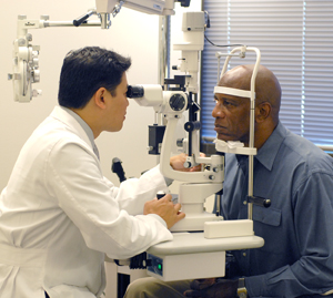
Slit lamp exam. This test looks at the structures of your eye.
-
Ultrasound. This makes an image of the eye using sound waves. It may be used if blood is found in the clear gel that fills the eye (vitreous).
-
Ocular coherence tomography. This test makes an image of the retina using light waves. This shows if there's fluid leaking into some parts of the eye. It can also measure how thick the retina is.
Fluorescein angiography
This test may be done to check the inside lining of the eye (retina). It also checks the tiny blood vessels (capillaries) that carry blood to the retina. During the test:
-
You'll be given drops that dilate your pupil.
-
A dye is then injected into the blood through your arm or hand. The dye travels to the capillaries in the eye.
-
The eye care provider takes photos of the retina. The dye makes the capillaries easy to see on the photos. This helps the provider know if and where there are problems.
You may feel nauseated for a short time during the test. For a few hours after the test, your skin, eyes, and urine may look yellow. Talk with your eye care provider to learn more about this test.